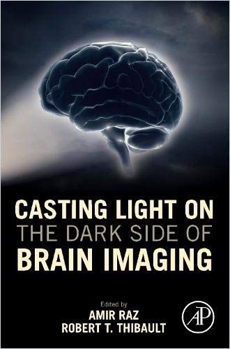
Neuroscientist Amir Raz and postdoctoral researcher Robert T. Thibault offer an overview of brain imaging technology to separate facts from popular perceptions and marketing hype.

Debunking Brain Claims
The human brain is a black box – stimuli go in, thoughts, words and precursors to behavior emerge. Nobody knows what happens in the interim. Then brain imaging technology and its accompanying hype appeared. And guess what? The brain remains a mysterious black box. So say Amir Raz – Canada Research Chair in the Cognitive Neuroscience of Attention at the department of psychiatry at McGill University and Robert T. Thibault – postdoctoral researcher at the University of Bristol. They assert that science and medicine know little about how the brain works, and that scientific evidence doesn’t support the roles people envision for brain imaging in psychiatry, medicine and even criminal law.
Brain Imaging
Electroencephalography (EEG) is a brain imaging technique. In noninvasive EEG, electrodes attached to the scalp record the electrical activity of brain cells. A related method, magnetoencephalography (MEG), records changes in magnetic fields the electrical activity of brain cells evoke.
Despite decades of research, we still don’t have a full understanding of how these signals are generated, what they represent, and whether they are a cause or effect of behavior.Amir Raz and Robert T. Thibault
Scientists use the data to understand how brain waves of various frequencies correlate to varied states of consciousness. EEG and MEG, the authors explain, allow scientists to observe phenomena that correlate to thought – not necessarily the phenomena that cause thought, and certainly not thought itself.
Scientists have difficulty discerning between brain activity and the smaller signals that originate in muscles. The “embodiment” model acknowledges that the brain doesn’t operate in isolation. It’s part of the body, which is part of the larger “social world.”
Functional Magnetic Resonance Imaging (fMRI)
Functional magnetic resonance imaging (fMRI) is a method for observing variations in “metabolism and blood flow” within the brain.
fMRI is used to infer changes in neuronal activity based on local changes in metabolism and blood flow. Such changes have been shown experimentally to accompany changes in neuronal activity, but the relationship is not necessarily one-to-one.Amir Raz and Robert T. Thibault
By observing indicators of blood flow, researchers glimpse which areas of the brain are most active. Brain activity varies by the millisecond – but fMRI detects changes only from second to second, displays brain signals about four to six seconds after they occur, and locates brain activity only within one millimeter.
fMRI is expensive, so studies tend to be “underpowered” – they have fewer participants and thus, the authors point out, less reliable results.
Research Replication Crisis
A scientific endeavor achieves credibility when a different set of scientists in a different place attempting to replicate published research obtain similar results. In 2015, for example, the field of psychology suffered a shake-up when scientists attempted to replicate 100 of the most important psychology studies, but obtained similar results only in 39. Brain imaging studies face barriers to proper replication because few researchers want to spend what brain imaging costs only to validate another researcher’s results.
But, the authors detail, researchers can offer “preregistration” – researchers explain their plan to a community of peers before conducting research. Clinicaltrials.gov and the open science framework (OSF) offer preregistration platforms. “Preprints” or “registered reports” allow peers to make suggestions or discuss research prior to publication.
Reproducing previous research mitigates some of the expense of data collection. A second research team reanalyzes existing data from a previous study to validate the study’s results. Original researchers must document their methods thoroughly, disclose relevant details, reveal data and share materials. Replication and reproduction, the authors affirm, improve the reliability and relevance of research findings.
“Unfalsifiable” Claims
Unfalsifiable claims don’t produce meaningful predictions, and cannot be falsified because they’re too nebulous to be refuted. The authors cite as an example the concept that addiction is a brain disease. This suggests that drug use alters the brain, and the resulting damage renders the drug user unable to withstand drug cravings.
Addicts’ capacity for choice-making, albeit at times compromised, is by no means obliterated in the face of demonstrable brain changes.Amir Raz and Robert T. Thibault
This idea took hold in the popular and professional consciousness. However, the authors say, the first part of the claim isn’t falsifiable, and science doesn’t support it. Drug use alters the brain, but every activity alters the brain, including juggling and reading.
Neuroimaging Roles
Brain imaging facilitates computer-assisted communication and the use of prostheses in people with paralysis, for example.
Researchers understand that to advance their ideas, they often must push their theories to the extreme. Popular media seldom temper their enthusiasm when reporting about brain imaging – thus misunderstanding, oversimplifying or hyping results. Self-ordained experts overstate the applicability of brain imaging and profit from doing so, the authors advise.
Conclusions drawn from neuroimaging assays have the potential to overpromise and underdeliver, and we can’t always be sure that advertising based on neuroscience presents a full story.Amir Raz and Robert T. Thibault
Purveyors of “brain-based” diagnostics, education, leadership plans, training and mindfulness programs take advantage of people’s enthusiasm for brain imaging – and their concerns about brain health – by marketing unsubstantiated claims about how their products benefit the brain. “Brain magic,” the authors warn, invades the courtroom in the form of “neurodeterminism” – the state of a person’s brain determines behavior. So if a brain scan reveals an abnormality, a person might not be responsible for his or her actions.
Heady Stuff
Raz and Thibault want to educate you in the field of brain imaging and all its wonders. They also want you to understand the limitations of brain imaging and to curb your enthusiasm regarding credible-sounding but false claims about brain imaging’s possibilities. To fully appreciate this education requires curiosity about the brain’s functions, brain imaging, scientific method, scientific cultural politics, popular media, the popular marketplace – and the many and surprising ways they intertwine. Raz and Thibault demonstrate their expertise on every page, with an astonishing lack of jargon. There are few scientific/medical and/or scientific/medical debunking treatises as readable as theirs. Some effort may be required when you first enter the portals of their universe, but the authors reward those efforts amply.
Amir Raz co-wrote How (Not) to Train the Brain with Sheida Rabipour; and Hypnosis and Meditation with Michael Lifshitz.


